Thyroid gland (TG) is the part of endocrine system
(the system of organs, producing biologically active substances (Hormones). Tissue of TG
secretes thyroid hormones (thyroxine (Т4) and triiodothyronine
(Т3)) into blood. TG hormones act on many different target cells in the body and
participate in great number of processes in organism: breathing, nutrition, sleep, movement,
and functioning of various organs (heart beating, reproductive system, etc.).
The thyroid gland is controlled by thyrotropin (TSH), secreted from the
pituitary gland, which, in turn, is influenced by the thyrotropin-releasing hormone
(TRH) from the hypothalamus. TSH permits growth, cellular differentiation, and thyroid
hormone production and secretion by the thyroid gland.
The most wide-spread diseases of TG are related to the lack of Iodine in food. According to
the World Health Organization data, diseases of thyroid gland are marked in 10-15 % of the
World population. More than 1 billion people live in the regions with iodine deficiency
(almost all the territory of Russia and continental Europe, central regions of Africa and
South America).
The most obvious manifestation of iodine deficiency in the body is endemic goiter
(diffuse enlargement of thyroid gland without dysfunction). Goiter formation is the
compensatory reaction directed on maintenance of the level of thyroid hormones in the body.
Insufficient iodine entry with nutrition causes change of thyroid gland function. Iodine
deficiency leads to reduction of synthesis and a secretion of Т4 and Т3 hormones for which
Iodine is "building material". It results in activation of thyrotropin (TSH) secretion.
Increasing of TSH concentration is one of indicators of iodine deficiency. Increased TSH
causes increased cellularity and hyperplasia of the thyroid gland in an attempt to normalize
thyroid hormone levels. If this process is sustained, a goiter is established. Endemic
goiter is contributing factor for development of many thyroid gland diseases, including
thyroid nodules and a cancer.
Treatment
Traditional methods of treatment are surgery (excision of pathological mass) and
conservative therapy (thyroid hormones). The disadvantages of these methods are low
efficiency, complications, relapses and trauma (in surgical treatment).
At present, low-invasive percutaneous interventions under ultrasonic control are used
for treatment of various thyroid diseases. They allow to eliminate the pathological focus
by influence of physical (laser) or chemical (ethanol sclerotherapy) factor,
preserving the major part of hormone producing thyroid tissue. Scientists of the Center
developed new method of treatment of thyroid gland diseases with high-intensity laser
radiation (interstitial laser thermotherapy).
The method of laser-induced thermotherapy is based on irreversible damage of pathological
cells and tissues under high temperature and absence of those damages in the healthy
surrounding tissues. This effect can be achieved by local tissue heating up to 43-45°С.
At this temperature pathological cells die, and healthy survive. This thermal field is
created with the help of near infrared (0,8-1,1 microns) laser radiation, delivered to the
pathological focus through a flexible fiber light guide. Advantages of this treatment are
locality and selectivity of action, the opportunity of treatment of deep structures, and
absence of serious complications.
Scheme and course of operation
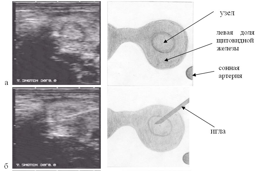
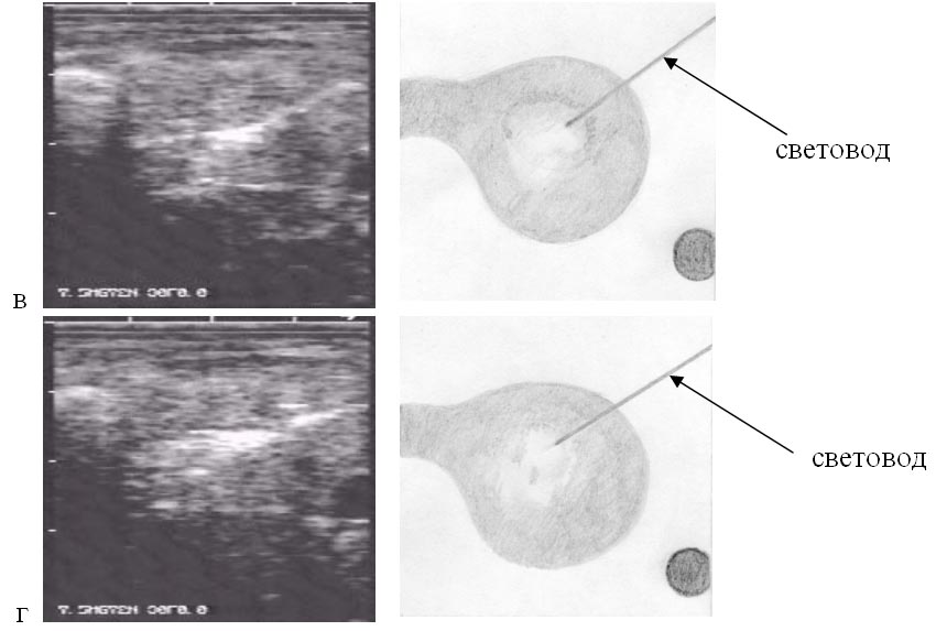
Fig. 1. Ultrasonic picture of laser interstitial thermotherapy of thyroid nodules.
a) heterogeneous node with inspissation in the center with hypoechoic border in the left lobe
of thyroid gland;
b) the aspiration needle punctured into the left lobe of thyroid gland;
c) the beginning of laser thermotherapy. Hyperechoic cloud in the zone of irradiation;
d) the end of laser thermotherapy. Hyperechoic zone without precise border in the projection
of the node.
Results
Чрескожная ЛИТТ-терапия с использованием диодных лазеров (0,81 и 1,06
мкм) под контролем УЗИ проведена нами у более 100 больных с различными видами
узлового зоба. Все они лечились амбулаторно и хорошо перенесли лазертермию.
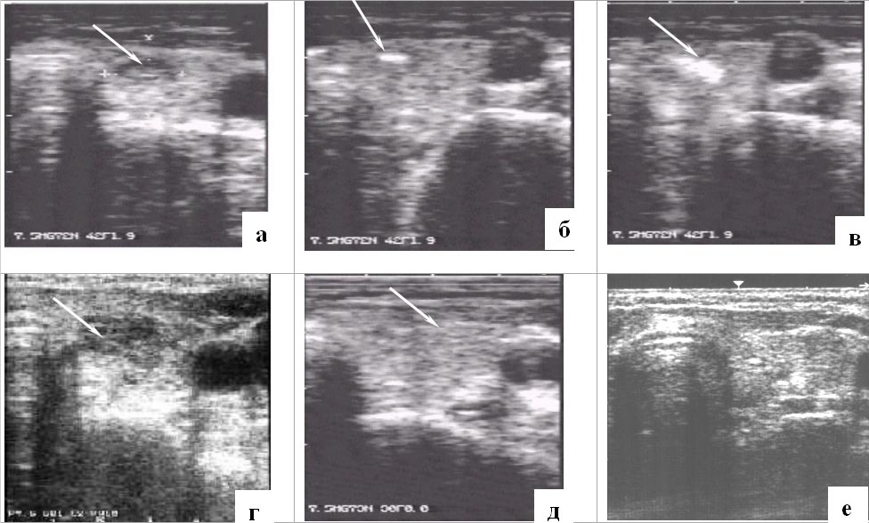
Hyperechoic cloud at the end of the lightguide was marked on the display of the ultrasonic
scanner in 0,5-1 mines after the beginning of the procedure (Fig. 2b). It gradually enlarged
and by the end of the laser thermotherapy "covered" the most part of the node (Fig. 2c).
Since the second day, the hypoechoic zone without precise borders (Fig. 2d) was formed
in the place of the node subjected to LITT. In 1-3 months after LITT the nonuniform
hypoecoic area with blurred borders and increasing focal fibrous changes was found in the
place of nodes (Fig. 2e). Later the node size decreased and fibrous changes developed in it
(Fig. 2f).
Fig. 2. Ultrasonic picture of nodular thyroid LITT;
a) Ultrasonography of the left lobe of thyroid gland. Heterogeneous node with hypoechoic rim
at the periphery;
b) the beginning of laser thermotherapy: hyperechoic cloud in the zone of irradiation;
c) the end of laser thermotherapy; hyperechoic zone without precise border in the projection
of the node;
d) the 2-d day after LITT: hypoechoic zone in the node projection;
e) 3 months after LITT: the node in the left lobe is not determined;
f) 1.5 years after LITT: the node is absent.
There were no complications during interstitial thermotherapy. From 1 to 6 procedures were
necessary depending on the node size. In 3-6 months after LITT, the gentle cicatrix without
deformation and changes in surrounding tissue was formed in the node place. Function of the
thyroid gland did not suffer (levels of TSH, Т3 and Т4 hormones did not change). Long-term
observation of 28 patients (1,5 - 3,5 years) showed good results in 93 % of them.
The nodes have decreased by more than 50%, or have not been revealed at all. Node growth was
marked in no case.
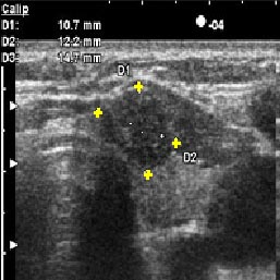
before treatment (nodular volume is 2.1 cm3)
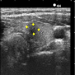
In 1,5 months after LITT (nodular volume is 0.6 cm3)
Fig. 3. Ultrasonic scanning images of the left lobe of thyroid gland with a node.